
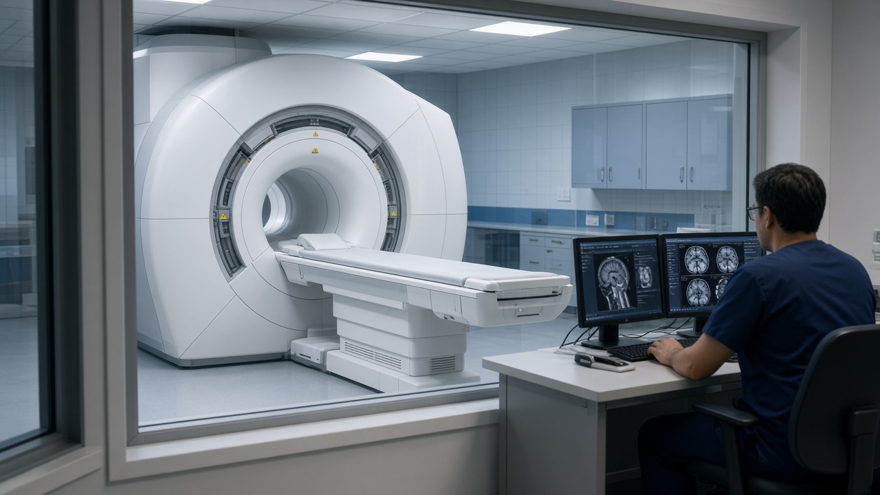
Start with water
Most of your body is water, and every water molecule contains hydrogen. The nucleus of a hydrogen atom behaves like a tiny magnet with its own spin. An MRI scanner works by nudging billions of these tiny magnets and listening to what they do next. No part of the process involves radiation.
Step one: the main magnet
The scanner is built around a large magnet that is always on, even when nobody is being scanned. When you lie in it, a small majority of your hydrogen nuclei line up with that field. Field strength is measured in Tesla; clinical scanners are usually 1.5T or 3.0T, which is thousands of times stronger than the earth's magnetic field. That is why metal is kept out of the room, and why the safety questionnaire matters.
Step two: the radio pulse
A coil sends a short radio pulse at exactly the frequency the aligned nuclei respond to, which tips them out of alignment. When the pulse stops they return to their original position and release the absorbed energy as a faint radio signal. Antennas in the coil placed around the body part being examined pick that signal up.
Step three: locating the signal
A signal on its own says nothing about where it came from. Gradient coils solve this by varying the magnetic field very slightly across the body, so the signal frequency differs from one position to the next. A computer works backwards from those frequencies to build an image. The gradient coils switch on and off many times a second, and the resulting vibration is the knocking noise you hear during the scan.
Why tissues look different
Different tissues release energy at different rates. Radiologists exploit this by running several sequences with different timings:
- T1-weighted images show anatomy clearly, with fat appearing bright.
- T2-weighted images make fluid bright, which highlights inflammation and swelling.
- Diffusion-weighted images track the movement of water molecules, useful in stroke and in tumours.
A single examination combines several of these. That is why an MRI takes 20 to 45 minutes rather than the few seconds of an X-ray: each sequence is a separate measurement, and the scan is complete only when the radiologist has the full set.
Where contrast fits in
Some questions need a contrast agent, usually based on gadolinium, injected into a vein. It changes how quickly nearby tissue releases its signal, making blood vessels, inflammation and many tumours stand out. Contrast is not used for every scan, and your clinic will tell you in advance whether your protocol needs it.
What this means for you in the scanner
Understanding the mechanism explains the two things patients are asked to do. Stay still, because the computer assumes the body has not moved between measurements. Accept the noise, because it is the sound of the gradients doing the work of locating each signal. Earplugs or headphones are always provided, and you can speak to the technologist between sequences.