
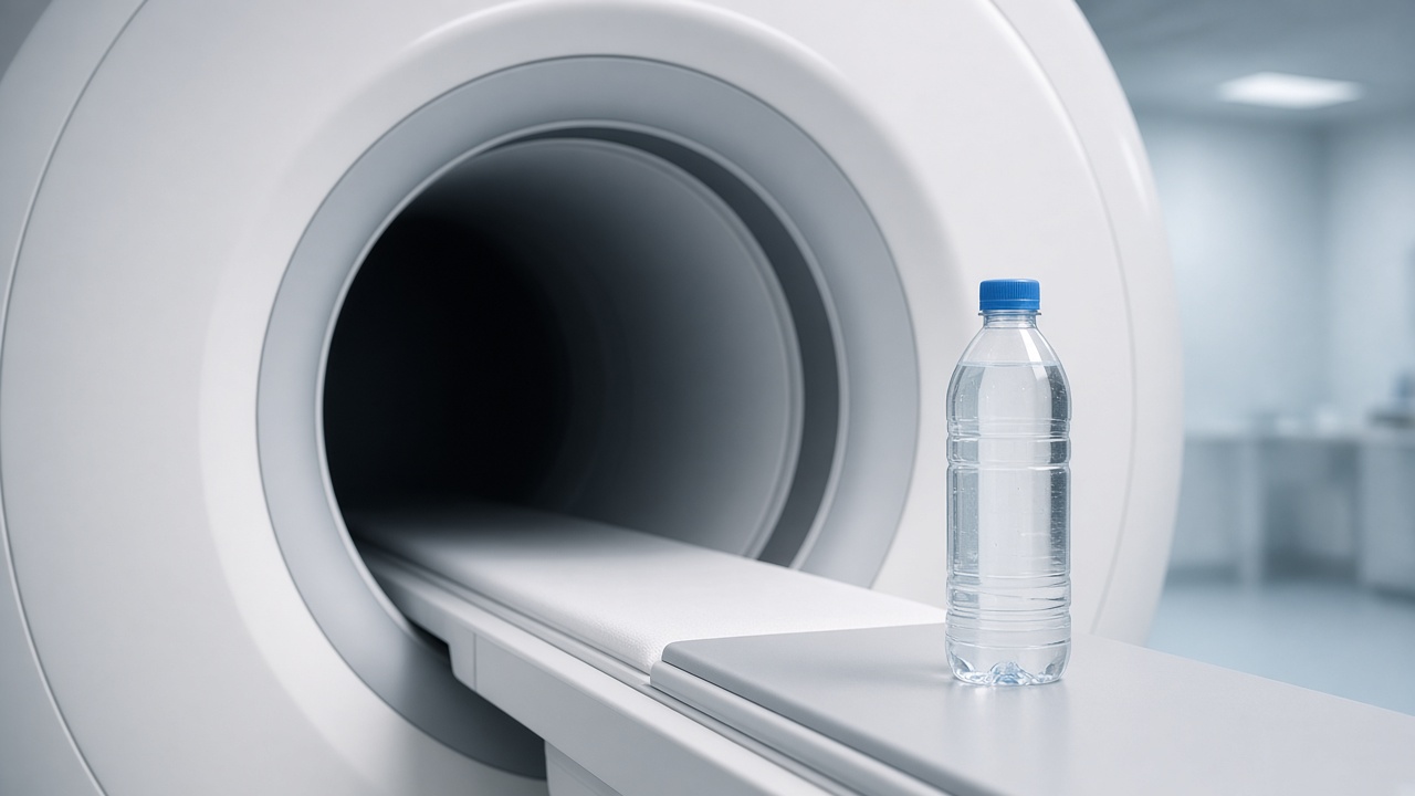
A magnet, not a camera
An MRI scanner does not photograph you. It uses a strong magnet to line up hydrogen atoms in water and fat. A short radio pulse tips them. When the pulse stops, they relax and give off a tiny signal. Coils around the part being scanned pick that signal up, and a computer turns it into slices.
Why the pictures look different from a CT
Different tissues relax at different speeds. That is why muscle, fluid, fat and many organs can be told apart. The radiologist chooses sequences — you will hear them as separate blocks of noise — so each block answers a different question. T1-weighted images emphasise anatomy. T2-weighted images emphasise fluid.
What you notice
You do not feel the magnet. You do hear the gradients, which sound like knocking. The table may move a few centimetres between sequences. Nothing is “running” through you. If contrast is used, that is a separate injection, explained in the contrast guides.