
Three tests, three jobs
Abdominal imaging usually starts with ultrasound, because it is quick, radiation-free and answers many questions on its own. CT is the workhorse for acute problems and for staging. MRI is the detail test: it characterises what the other two have found, particularly in the liver, bile ducts and pancreas.
What abdominal MRI is used for
- Liver lesions — distinguishing a harmless haemangioma or area of focal fat from something that needs treatment.
- Bile ducts and gallbladder — MRCP images the duct system without a catheter and shows stones and strictures.
- Pancreas — cystic lesions, chronic inflammation and suspected tumours.
- Liver fat and iron — modern sequences quantify both without a biopsy, which is used to monitor fatty liver disease and iron overload.
- Kidneys and adrenal glands — characterising a mass found incidentally on another scan.
- Bowel — MR enterography follows Crohn's disease over years without repeated radiation.
Why not simply use CT?
CT is faster and better for gas, calcification and acute bleeding, and it remains first choice in an emergency. It does, however, involve ionising radiation, and it cannot always separate one soft-tissue density from another. MRI's several contrast mechanisms let a radiologist say what a lesion is made of — fat, fluid, blood or solid tissue — which is often the decisive question. For patients who need repeated follow-up, avoiding cumulative radiation is a genuine advantage.
Why not simply use ultrasound?
Ultrasound is excellent for gallstones, obstructed kidneys and a first look at the liver, at a fraction of the cost of MRI. Its limits are physical: bowel gas and body build can obscure the pancreas and other deep structures, and image quality depends heavily on the operator. When ultrasound finds something it cannot fully characterise, MRI is often the logical next step.
Preparation and the appointment
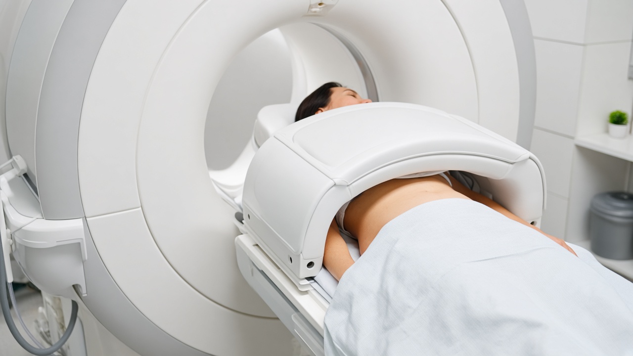
Abdominal MRI usually requires fasting for four to six hours, so that the gallbladder is distended and the stomach empty. Contrast is common, and some liver protocols use a liver-specific agent that requires a delayed series around 20 minutes after injection. The examination takes 30 to 45 minutes.
Breath-holding is the part patients find demanding. Many sequences need you to hold your breath for 15 to 20 seconds at a time so that movement does not blur the image. The technologist coaches you through each one, and free-breathing alternatives exist if you cannot manage it.
What happens with the result
A written report goes to the doctor who referred you, who interprets it alongside your symptoms and blood tests. Incidental findings such as small liver or kidney cysts are very common and usually need nothing beyond reassurance. Any decision about follow-up imaging, biopsy or treatment belongs with your specialist.