
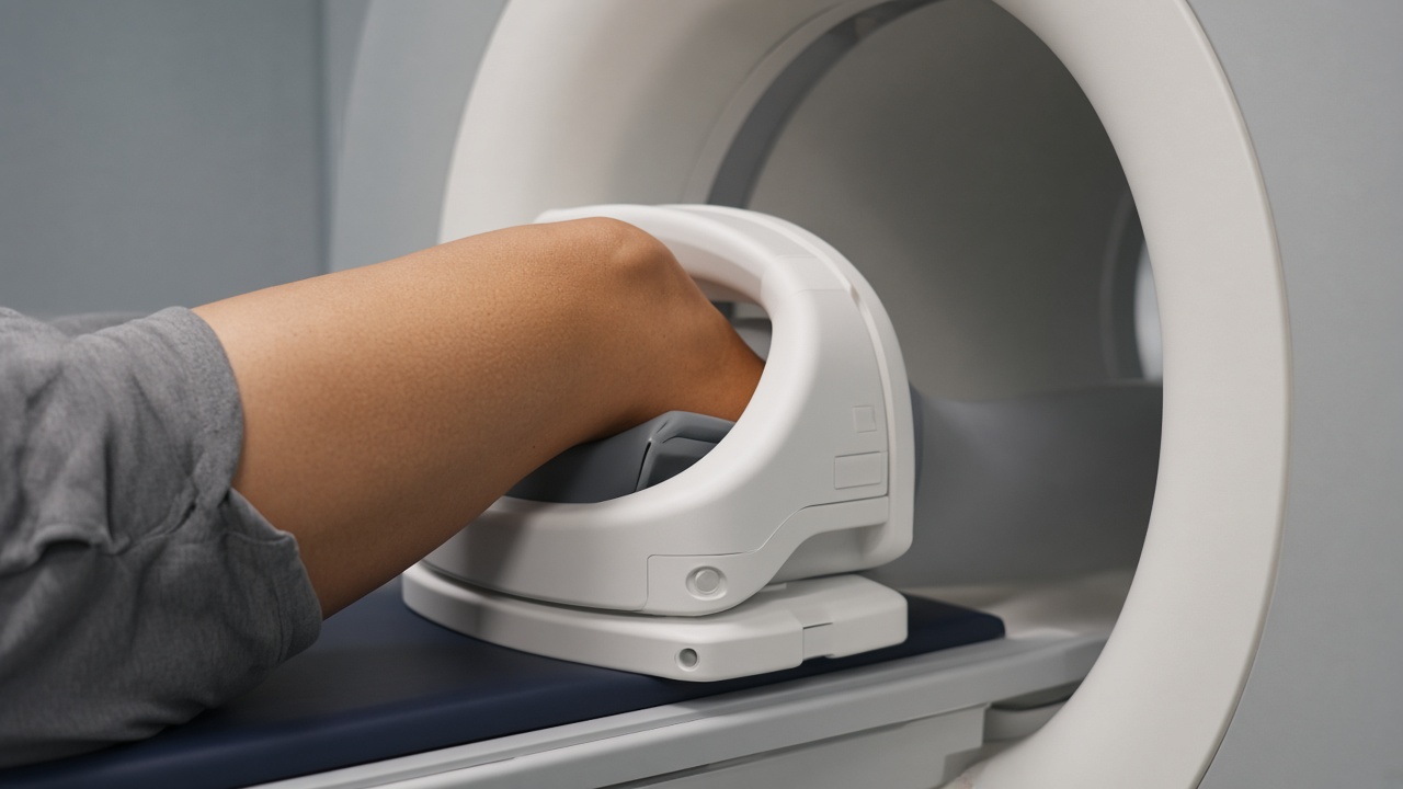
Why joints are an MRI strength
X-ray shows bone. Ultrasound shows structures close to the surface. MRI shows the soft tissue inside a joint — cartilage, ligaments, tendons and the joint lining — in a single examination, which is why it is the standard test when injury inside a joint is suspected.
Knee MRI
A knee protocol is most often requested after a twisting injury, or when the joint locks, gives way or swells repeatedly. It reliably shows:
- Meniscal tears — including the type and position of the tear.
- Cruciate and collateral ligaments — partial versus complete tears.
- Cartilage damage — early loss of cartilage, before it becomes visible on X-ray.
- Bone bruising and stress fractures — often invisible on plain films.
- Fluid, cysts and inflammation — such as a Baker's cyst behind the knee.
Shoulder MRI
The shoulder is a shallow joint held together by tendons and a rim of cartilage, so soft-tissue detail is decisive. MRI is used for:
- Rotator cuff tendons — tendinopathy, partial tears and full-thickness tears, including the size of the gap.
- Labral tears — after dislocation, or in throwing athletes.
- Impingement — bursitis and the shape of the overlying bone.
- Frozen shoulder — thickening of the joint capsule.
For some shoulder questions, contrast is injected into the joint itself before scanning. This arthrogram technique separates structures and improves detection of small labral tears. It is a minor procedure performed by a radiologist and is used only when the clinical question requires it.
When ultrasound or X-ray comes first
For a superficial tendon problem, ultrasound is quick, inexpensive and allows the joint to be moved during the examination, which MRI cannot do. For a suspected fracture, established osteoarthritis or alignment questions, X-ray answers the question at a fraction of the cost. MRI is the right next step when the clinical picture suggests damage inside the joint, when surgery is being considered, or when simpler tests do not explain the symptoms.
Practical points
Joint scans are among the more comfortable MRI examinations. For the knee, only the affected leg needs to be inside the scanner, so your head usually stays outside the bore — useful if you are claustrophobic. A knee or shoulder protocol takes about 20 to 30 minutes. For the shoulder you lie with the arm at your side in a fixed position, which is the part patients find most tiring; tell the technologist if you cannot hold it.
Reading the report
MRI is sensitive enough to find changes in joints that do not hurt, particularly in people over 40. A degenerative meniscal tear or a worn rotator cuff tendon is a common finding at that age and does not automatically call for surgery. Your orthopaedic surgeon or sports physician weighs the images against the physical examination and how the joint actually functions.