
What it shows
A cardiac MRI looks at the heart muscle, the size of the chambers, some valve problems, and areas of scar or inflammation. It is used for cardiomyopathy, myocarditis, congenital heart disease and to sort out an unclear echocardiogram.
What it does not replace
- An ECG or blood tests in sudden chest pain.
- A coronary angiogram when doctors need to see and treat a blocked artery immediately.
- A simple echocardiogram, which is often enough and does not need a tunnel scanner.
What the appointment is like
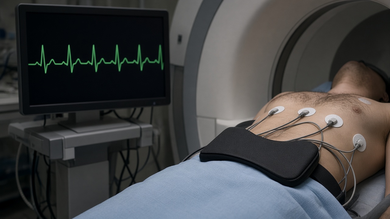
You lie still with ECG leads on your chest. The scanner takes pictures between heartbeats, so the exam is often longer than a knee MRI, commonly 45–60 minutes. Breath-holds are short and coached. Some studies use gadolinium to show scar. Tell the team about kidney disease and any pacemaker before you attend.