
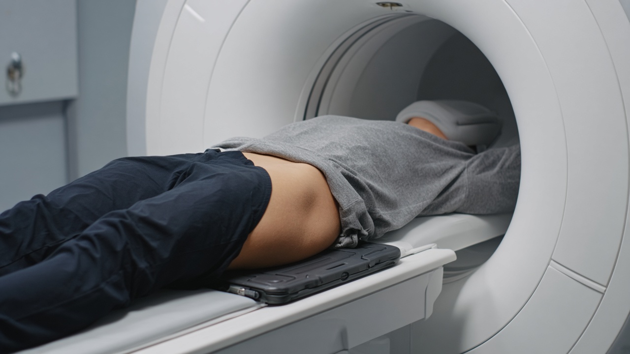
What a spine MRI looks at
An MRI of the spine shows the vertebrae, the discs between them, the spinal cord and the nerve roots leaving the spinal canal. It is the only routine test that displays all of these at once, which is why it is the standard examination when back or neck pain comes with nerve symptoms.
Common findings
- Disc herniation — whether disc material is pressing on a nerve root, and at which level.
- Spinal canal narrowing — degenerative stenosis that causes leg pain on walking.
- Wear and tear — dehydrated discs, facet joint arthrosis and bone marrow changes.
- Inflammation — sacroiliitis and other changes seen in inflammatory back disease.
- Fractures — including whether a collapse is recent or old, which X-ray cannot always tell.
- Tumours and infection — uncommon, but important to exclude when warning signs are present.
When a scan is not the answer
Most episodes of back pain settle within weeks without imaging. Dutch and international guidelines advise against early MRI for uncomplicated low back pain, for a straightforward reason: degenerative changes are found in a large share of people who have no pain at all. Scanning too early often produces findings that are real on the image but are not the cause of the problem, which can lead to unnecessary worry and treatment.
Imaging becomes appropriate when pain radiates into the leg or arm in a nerve pattern, when there is weakness or loss of sensation, when symptoms persist beyond six to eight weeks of conservative treatment, or when there are warning signs such as unexplained weight loss, fever, a history of cancer, or problems with bladder or bowel control. That last group needs urgent assessment rather than a routine appointment.
Which part of the spine?
Protocols are usually ordered by region: cervical (neck), thoracic (mid-back) or lumbar (lower back). Each takes roughly 20 to 30 minutes. A scan of the whole spine takes considerably longer and is reserved for questions that genuinely span all levels. Leg symptoms point to the lumbar region; arm symptoms point to the neck.
What to expect
You lie on your back, often with a cushion under the knees to flatten the lower spine and make the position easier to hold. Contrast is not routine, but it is used after previous spine surgery, or when infection or a tumour is suspected. Some clinics offer upright or weight-bearing MRI; this can add information in selected cases, but conventional MRI remains the reference standard.
After the scan
A radiologist correlates the images with your symptoms, because the level seen on the scan has to match the level of your complaints for a finding to be meaningful. Decisions about physiotherapy, injections or surgery are made by your clinician on the basis of the physical examination and the report together.