
Why MRI is the first choice for the brain
MRI gives the most detailed view of brain tissue of any routine imaging test. It separates grey matter from white matter, shows the fluid spaces clearly, and picks up changes that a CT scan can miss entirely. Because MRI uses a magnetic field and radio waves rather than X-rays, there is no radiation dose, which matters when scans have to be repeated over many years.
What a brain MRI can show
- Tumours and other masses — location, size and the relationship to surrounding structures.
- Multiple sclerosis — inflammatory lesions in white matter, and whether they are new or long-standing.
- Stroke — diffusion-weighted sequences show affected tissue within minutes of onset.
- Blood vessels — aneurysms and malformations, often with a dedicated angiography sequence.
- Memory problems — patterns of shrinkage and small-vessel change that help characterise dementia.
- Pituitary gland and inner ear — small structures that need thin slices and high resolution.
When CT is used instead
In an emergency, CT usually comes first. It takes seconds rather than half an hour, works with agitated or unstable patients, and reliably shows fresh bleeding and skull fractures. MRI follows once the situation is stable and a more detailed answer is needed. For questions about the sinuses and the bones of the face, CT also remains the better test.
Symptoms that often lead to a brain MRI
Referrers commonly request brain imaging for headaches with unusual features, new seizures, persistent dizziness or one-sided hearing loss, unexplained weakness or numbness, visual disturbance, or changes in memory and behaviour. Headache on its own, with no other findings, frequently does not require a scan at all.
What the scan involves
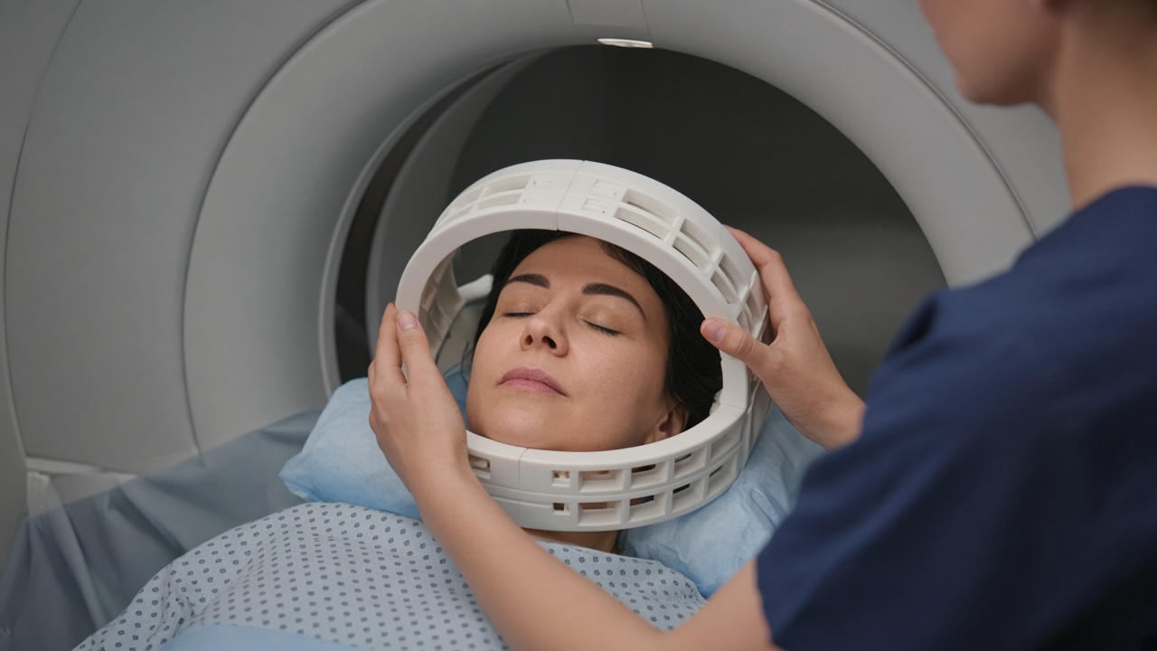
You lie on your back with your head resting in a padded coil, which sits close to your face without touching it. A brain protocol usually takes 20 to 40 minutes and is built from several sequences of a few minutes each. Contrast is not always needed; it is added when the question involves a tumour, an infection or active inflammation.
Staying still matters more for the head than for almost any other region, because movement of a few millimetres blurs fine detail. If you expect to find the coil confining, say so in advance. Many clinics offer a wide-bore scanner, a mirror that lets you see out of the bore, or light sedation.
Interpreting the result
Brain MRI often shows small incidental findings with no clinical meaning, such as tiny cysts or minor white-matter spots that become more common with age. A radiologist reports these in context, and your doctor decides whether anything needs follow-up. Discuss the report with the clinician who referred you rather than drawing conclusions from the images yourself.